Unlocking the Secrets of Lifelong Health
Human health stems from a combination of countless factors, from genetic and environmental influences to all the patterns, habits and behaviors we engage in from day to day.
Many of those habits and behaviors take root in early childhood and follow us throughout our lives, adding up to a massive impact on our long-term health.
Sheila Crowell, a professor of psychology in the College of Arts and Sciences (CAS), wants to trace one of our most essential patterns for lifelong health—our ability to get quality sleep—back to its origins. She’s studying how sleep habits are formed and how they affect the brain development of babies, beginning in the womb.
“I think a lot of psychologists would say that much of who we are is about our habitual patterns, thoughts and behaviors, so we’re trying to get to the roots of how some of those very first habits around sleep are developing,” she says. “I think for a lot of reasons it’s one of hardest skills to learn. It you talk to any adult, a significant number of them have some issues around sleep, and a lot of that is developing in that first year of life.”
From the impact of sleep on developing infants to the link between artery stiffness and age-related memory disorders such as Alzheimer’s disease, faculty across CAS are engaged in a broad range of research aimed at improving health and wellness across the entire human lifespan.
While some are contributing to our fundamental knowledge in areas such as biology and human physiology, others are teasing out the causes behind specific disorders and even testing experimental treatments designed to make our lives better—and longer.

Tracing the Impact of Infant Sleep Patterns
Ask any new parent, and they’ll tell you lack of sleep is one of the biggest challenges they face during the first year of their baby’s life.
What’s less understood is how that lack of sleep during the first months of parenthood might affect a baby’s ability to develop healthy sleeping habits—and how those habits affect the child’s neurodevelopment.
“We know that across the lifespan, when people don’t sleep well, they’re more vulnerable to dysregulated emotions, difficulties with attention and focus, and they experience more behavioral problems,” says psychology Professor Sheila Crowell. “If this is true across the rest of the lifespan, it’s possible that it’s also true during that first year of life when a lot of brain development is happening.”
Crowell received a $4,025,596 grant from the National Institutes of Health to study the relationships between prenatal exposures, neurological development and sleep in infants—and how they affect behavior and emotional regulation in toddlers.
While previous studies have explored the link between sleep and problems with attention, behavior and emotional regulation in older children, none have focused on new babies, she adds.
“One of the things we’re curious about is to what extent do the birth parent’s own biological rhythms and stress around sleep affect differences in how the baby is born?” says Crowell, who is working in collaboration with Liz Conradt, an associate professor at Duke University. “And what about their partner? If they’re having disrupted sleep or different sleep hours, how does that affect the pregnant person and the baby?”
Another novel aspect of the study is that it will examine the family’s sleep patterns before the baby is born, beginning during the second trimester of pregnancy—a sensitive time for brain development. Parents will complete surveys and engage in periods of sleep tracking during both the second and third trimesters.
“We’re interested in what’s happening with the whole family system and how that affects the baby,” Crowell says.
Once the babies are born, Crowell’s team will administer an assessment known as the NICU Network Behavioral Scale (NNNS), which measures an infant’s neurological and behavioral development at birth.
“We suspect we’ll see signs of differences in attention and behavior already in the first 24 hours after birth, and those differences, along with the sleep routines in the family, will help us to see which kids are developing skills for sleeping early and which ones are taking longer to develop those skills,” Crowell says.
The study will follow participants throughout the first year of their babies’ lives, with Fitbits to track the parents’ sleep patterns and Owlet Socks for monitoring the babies’ heart rate and oxygen levels to determine when and how much they sleep.
“We have all these capabilities that allow us to look at their attention and emotional regulation at birth and see already how their brains are working a bit, so we can then look at the extent to which that’s affecting their sleep and whether it’s affecting their attention profiles at one year,” Crowell says.
She hopes the study will help combat misinformation and provide parents with reliable guidance on the natural range of infant sleeping skills, as well as what factors might affect a baby's ability to self-soothe.
“We all know that sleep is challenging in the first year of life and that the baby is developing its first skills for figuring out how to self-soothe, fall asleep and stay asleep,” Crowell says. “Because there aren’t clear guidelines for what the first year of sleep is going to look like, the parent feels alone. They don’t know if the baby is sleeping enough or too much.
“Our goal really is to empower parents to understand what types of things might help their baby sleep more effectively.”

Sheila Crowell
Psychology: Sleep and neurodevelopment
Received $4,025,596 from the National Institutes of Health to study the relationships between prenatal exposures, neurological development and sleep in infants and how they affect behavior and emotional regulation in toddlers
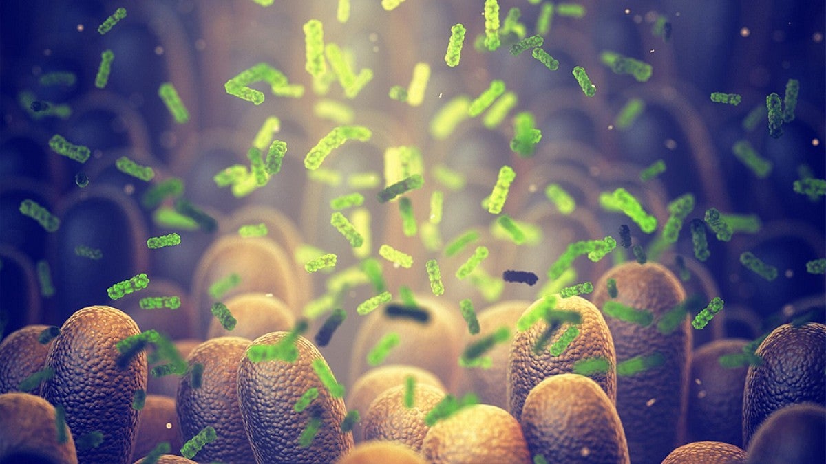
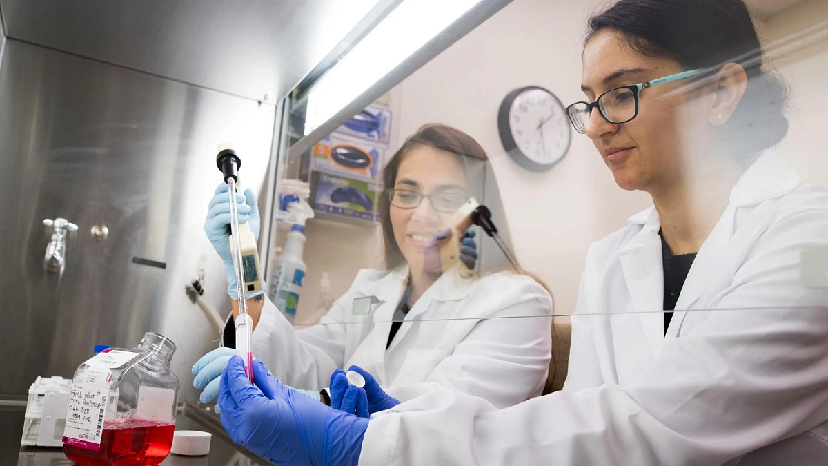
Understanding How the Gut Microbiome Works
Your gut microbiome—the complex ecosystem of bacteria that live in your intestines—plays a vital role in your long-term health, affecting everything from nutrient absorption to your immune system. When this system becomes imbalanced, it can contribute to diseases such as diabetes and certain cancers.
But there’s a lot that scientists still don’t know about how it works.
Romila Mascarenhas, assistant professor of chemistry and biochemistry, is contributing to our fundamental understanding of the gut microbiome by studying how bacteria acquire and use vitamin B12.
Vitamin B12 is an essential nutrient for a range of bodily functions such as energy production and DNA synthesis. We can’t survive without it, yet our bodies aren’t able to produce it.
Our gut bacteria also need it. Of the trillions of microbes living in the gut, 90 percent require B12 for survival, but only about 50 percent are able to synthesize it on their own. The rest rely on their environment to provide it.
“The gut is a competitive environment,” says Mascarenhas, who received $747,000 from the National Institutes of Health to study vitamin B12 trafficking in gut bacteria.
“They have complex B12 trafficking pathways and encode B12-binding proteins involved in scavenging this precious cofactor. Understanding how B12 is imported and shared between different gut microbes is important for understanding how these communities compete for or share their nutrients.”
Bacteria express specific membrane proteins that serve as transporters to import nutrients. Her lab is exploring the molecular mechanisms involved in transporting vitamin B12, as well as the relationships between microbes in the gut.
“Elucidating the molecular mechanics of vitamin B12 trafficking and selectivity will provide insights into how bacteria compete with each other or their host within this very complex ecosystem,” she says.
When harmful bacteria flourish and helpful bacteria are suppressed, the gut microbiome becomes imbalanced, which can contribute to a broad range of diseases. With a deeper understanding of how this ecosystem works, doctors could potentially help treat or prevent diseases by altering the composition of the microbiome, Mascarenhas says.
“Understanding these mechanics will allow us to develop strategies for targeted manipulation of the gut microbiome. For example, if we wanted to target a specific microbe in the biome, we can use B12 as a model for understanding its nutrient acquisition strategy.”

Romila Mascarenhas
Chemistry and Biochemistry: Gut microbiome
Received $747,000 from the National Institutes of Health to study how bacteria in the intestines acquire and use vitamin B12.
Exploring the Neuroscience of Movement
Imagine walking on campus. You get to your building and stop because you’ve arrived at your destination. Now imagine you’re on your way to your building, but you stop suddenly because you see someone coming at you on a skateboard.
In both examples, you stop moving. But what happens in your brain is different.
Understanding brain activity during the termination of movement in these different situations could play a key role in better understanding a broad range of disorders, from Parkinson's disease to obsessive-compulsive disorder (OCD), says Nicki Swann, assistant professor of human physiology.
With a $2.8 million grant from the National Institutes of Health, she’s using a combination of brain stimulation and brain recordings in humans to see what’s happening when we stop moving in different situations.
Her research focuses on the cortical basal ganglia network, a complex system of brain circuits that plays a key role in motor control, cognitive function and emotional regulation. Many different disorders affect this region of the brain, including OCD, ADHD, Parkinson’s disease and substance abuse.
Although movement is largely a motor function involving the physical movement of muscles, it also has a cognitive component, Swann says.
“Imagine you’re about to cross the street and suddenly see a car zipping around the corner, and you stop yourself,” she says. “The act of stopping yourself recruits a cognitive brain circuit that seems to be altered in a number of disorders.”
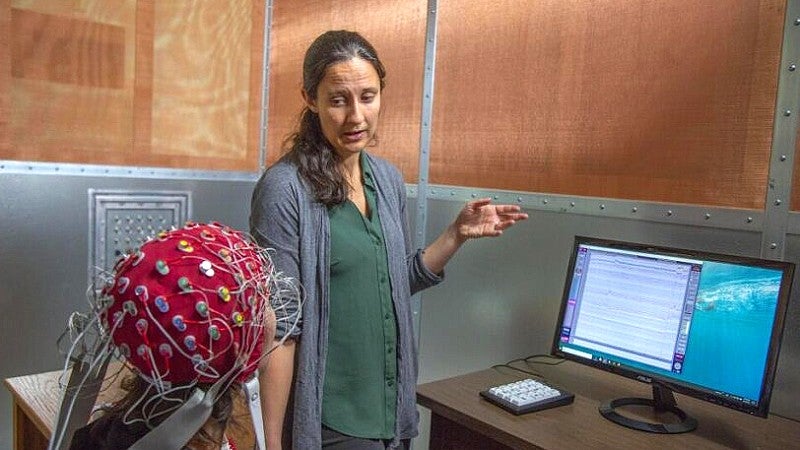
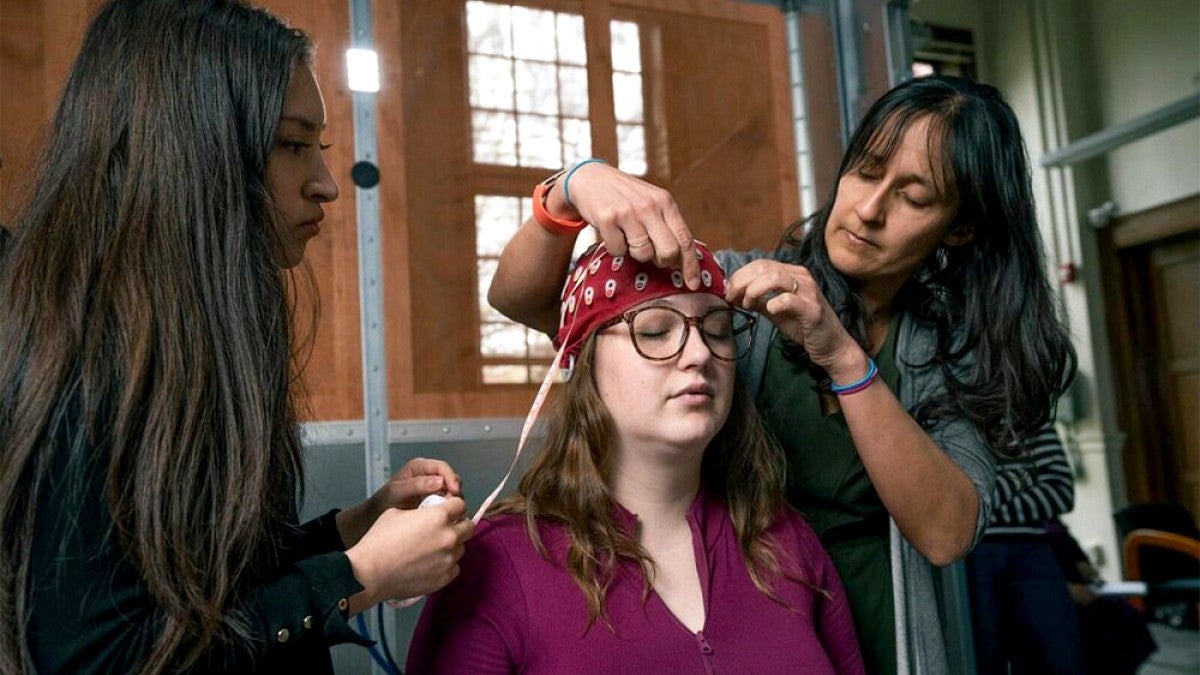
Swann’s team has designed their own research task in which subjects engage in continuous movement and then stop themselves in different contexts. Throughout the task, Swann’s research team monitors the subject’s brain activity using either electroencephalogram (EEG) or intracranial EEG to look for patterns in the brain’s electrical activity that occur when the movement stops.
“This test allows us to tease out the motor and cognitive aspects and pull apart behaviors that were previously intertwined,” she says. “During this movement task we can examine what’s going on in the cognitive part of the brain.”
Her team, which includes research associate Sara Parmigiani as well as PhD students Apoorva Karekal, Kelsey Schultz, Brittany Lyon and Shahrzad Ayoubipour and undergrad Cinthia Muñiz Sanchez, is collaborating with surgeons at OHSU to study the brain activity of patients with electrode implants, sometimes used to treat epilepsy, which yield more detailed data than a typical EEG.
Their research has the potential to improve treatment for disorders such as Parkinson’s disease. By understanding which signatures in brain activity correspond to improved or worsening symptoms, doctors could potentially use EEG readings to directly inform clinical care.
“We don’t currently have an objective way to quantify the severity of Parkinson’s disease, and sometimes that can be a real barrier,” Swann says, adding that clinicians rely on physical exams and patient self-reporting, both based on subjective rating scales, to diagnose and monitor the disorder.
“There’s a push in the field to see if we can identify objective ways to diagnose and assess severity to determine when it’s time to adjust patients' medication. Can we see signals that there is progression before the patient sees symptoms, and intervene with a higher dose of medication or therapeutic brain stimulation?”

Nicki Swann
Human Physiology: Neuroscience of movement
Received $2.8 million from the National Institutes of Health to study what happens in the brain when we stop moving.
Examining the link between blood vessels and Alzheimer’s
Our blood vessels stiffen as we age, making it harder for blood to flow through the arteries. This can increase the risk of heart disease, but it’s also correlated with Alzheimer’s disease, dementia and other memory disorders associated with aging.
What scientists don’t know yet is why.
Ashley Walker, associate professor of human physiology, has spent the past several years studying this correlation with funding from the National Institutes of Health. “We really want to get at the causative nature of what’s happening with the blood vessels and how it’s impacting the brain,” she says.
Her $2.1 million grant funds the study of mice that have been genetically modified to remove one of the proteins responsible for blood vessel elasticity, which does not negatively affect the welfare of the mice but allows researchers to study how the lack of elasticity affects their brain functions. In collaboration with OHSU, which has a magnetic resonance imaging (MRI) machine for small animals, she’s using MRI to examine the blood flow in their brains.
“We think the dysfunctional arteries are lowering the blood flow to the brain, which can starve tissue of oxygen and nutrients and lead to problems with memory,” Walker says.
She’s also working with a neuropathologist at OHSU to examine cell changes, inflammation and amyloid plaques (protein deposits) in the brain—a hallmark feature of Alzheimer’s disease.
While previous studies on artery stiffness have largely focused on male subjects, Walker is studying both males and females. Her team has found notable differences between the sexes in terms of how they respond to arterial stiffness and at what age they are affected.
Premenopausal females have high estrogen levels, which provide protection against cardiovascular disease and Alzheimer’s. When they enter perimenopause, however, their estrogen levels drop, their hormonal profile changes dramatically and their risk of these diseases shoots up. The risk of Alzheimer’s, for example, is twice as high for women as for men.
"Something about the menopause transition period, with the rapid change in hormones, is causing the risk to steeply incline,” Walker says. “Males don’t have that dramatic change to hormones with advancing age. We think that what’s partially causing the increased disease risk in females is the arteries not having time to adapt. We’re working on better understanding what’s happening during the menopause transition.”
Her research could help inform treatments for post-menopausal women, such as hormone replacement therapy and other potential therapeutics.
Walker has also explored potential interventions for Alzheimer’s disease, the seventh-leading cause of death in the US. In a previous study, she found that pyridoxamine, a form of vitamin B6, can help prevent stiffening arteries, reduce blood pressure, improve blood vessel function in the brain, and prevent some memory problems.
“There are no treatments to slow down, prevent or stop this disease,” Walker says. “This is an area we really need to figure out as the population gets older and the number of people at risk continues to grow.”

Ashley Walker
Human physiology: Blood vessel stiffening and Alzheimer's
Received $2.1 million to explore the link between stiffening blood vessels and age-related memory disorders such as Alzheimer's disease.
—By Nicole Krueger, College of Arts and Sciences