Experiential Learning | Research & Innovation | Community Impact | Career Preparation | Teaching Excellence | 21st Century Liberal Arts | Building Community | Good Vibes | CAS Spotlights | All Stories | Past Issues
A Window to the Brain
BY NICOLE KRUEGER
JUNE 9, 2025
For decades, scientists have believed that the outermost layer of our brain is divided into distinct areas, each responsible for performing different, separate information processing tasks.
But recent research suggests the reality is more complex than that—and Evan D. Vickers wants to find out why.
Using a novel technique he developed in Professor David A. McCormick’s neuroscience lab, Vickers is studying how the brain’s various regions react during different states of arousal, such as sleep versus wakefulness, both at the individual cell level and across large swathes of the cerebral cortex.
“I wanted to image all across the brain, in as many different areas as possible,” says Vickers, a research associate and postdoctoral scientist in the Institute of Neuroscience. “We’re trying to explain the different, distributed patterns of activity we see across the cortex related to arousal and movement.”
While scientists such as Vickers have studied the mechanisms of individual synapses (the electrical impulses nerve cells use to communicate with each other), less is understood about how neurons across the brain work in tandem during complex tasks such as decision making.
“Now we’re also looking at the big picture. How is the whole organ working?” he says. “I think we have to look at both at the same time.”
The cortex—the outermost layer of a mammal’s brain, responsible for higher-level cognitive functions like thinking and sensory processing—contains tens of thousands to millions of nerve cells, or neurons, clustered into roughly 60 different areas across both hemispheres. For example, there are different areas for visual processing, auditory processing and motor functions.
Using mice as a model, Vickers is trying to figure out why certain movements and arousal states trigger activity in neurons in multiple areas of the brain while others drive activity that is localized to a particular area.
For example, “in the dorsal [top] part of the brain, a bunch of cells turn on when the subject becomes active, and a bunch of cells turn off, and they’re all mixed together. But on the side of the brain, which contains auditory and somatosensory/touch processing and other associated areas, we see more localized patterns where all the cells that turn on or off following a change in arousal or movement are living in same area and keeping the other guys out.”
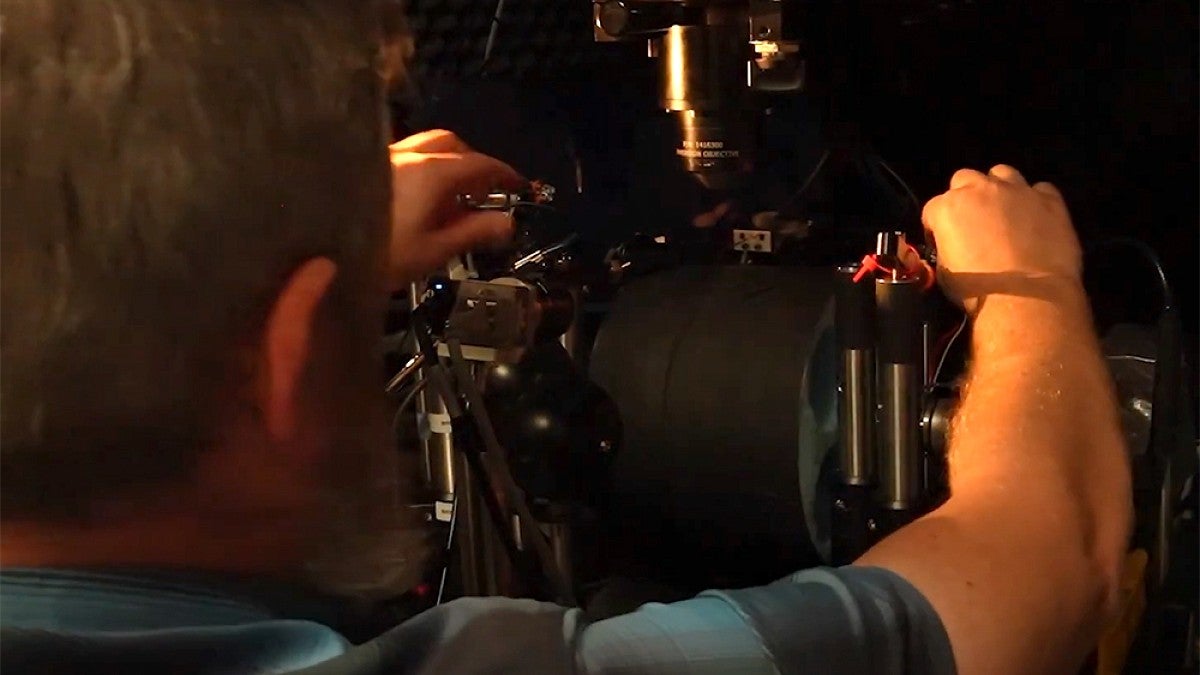
To determine which neurons “turn on” during different arousal states, Vickers uses two-photon microscopy, whose infrared laser pulses penetrate deeper into brain tissue than a typical microscope. But the standard version of this method can only image hundreds of neurons at a time—a drop in the bucket compared to the 15 million neurons in a mouse’s cortex.
To get to the bottom of what's really going on, Vickers needed to be able to examine a larger subset of neurons across multiple areas of the cortex.
He designed a special implant that replaces the entire skull with curved glass, creating a “window” to the brain. Together with a specialized two-photon microscope called a “mesoscope,” this cortical window allows researchers to image 10,000 or more neurons at a time without harming the animals, which live normal, pain-free lives.
Once the window is implanted, the mouse is either allowed to behave spontaneously on a running wheel or to perform a variety of decision-making tasks while responding to auditory or visual stimuli for one to two hours at a time. The microscope records its neural activity as the mouse transitions between different arousal states, which are measured as changes in pupil diameter, and performs different movements, such as walking or moving its whiskers.
In addition to dramatically expanding the number of cells that can be imaged at a time, the cortical window allows researchers to monitor neurons across multiple areas of the brain at once.
“Visual processing is in the back of the brain, and decision making is primarily in the front. We can now look at both at the same time, for example,” says Vickers, who published his technique in an eLife paper last year. “I think there’s a huge advantage to being able to do that.”
Equipped with this novel methodology, Vickers is now examining neural activity in other arousal states, such as when mice are under the influence of psychedelics. He also hopes other scientists will be able to use the large volume of data he's collecting for their own research.
“The field of psychedelics is sort of hot right now, and a lot of people are doing experiments,” he says. “But something that's missing is experiments where people have huge neural recordings like the kind that we do here, recording from tens of thousands of neurons at once.
“It's kind of cool to think about a new era of neuroscience where you have groups that are producing huge amounts of data and then you also have many groups that are all analyzing that data and looking for different patterns, in a coordinated team effort that will drive rapid, profound progress in the field.”
For any concerns regarding animal welfare, please visit the University of Oregon Institutional Animal Care and Use Committee.